
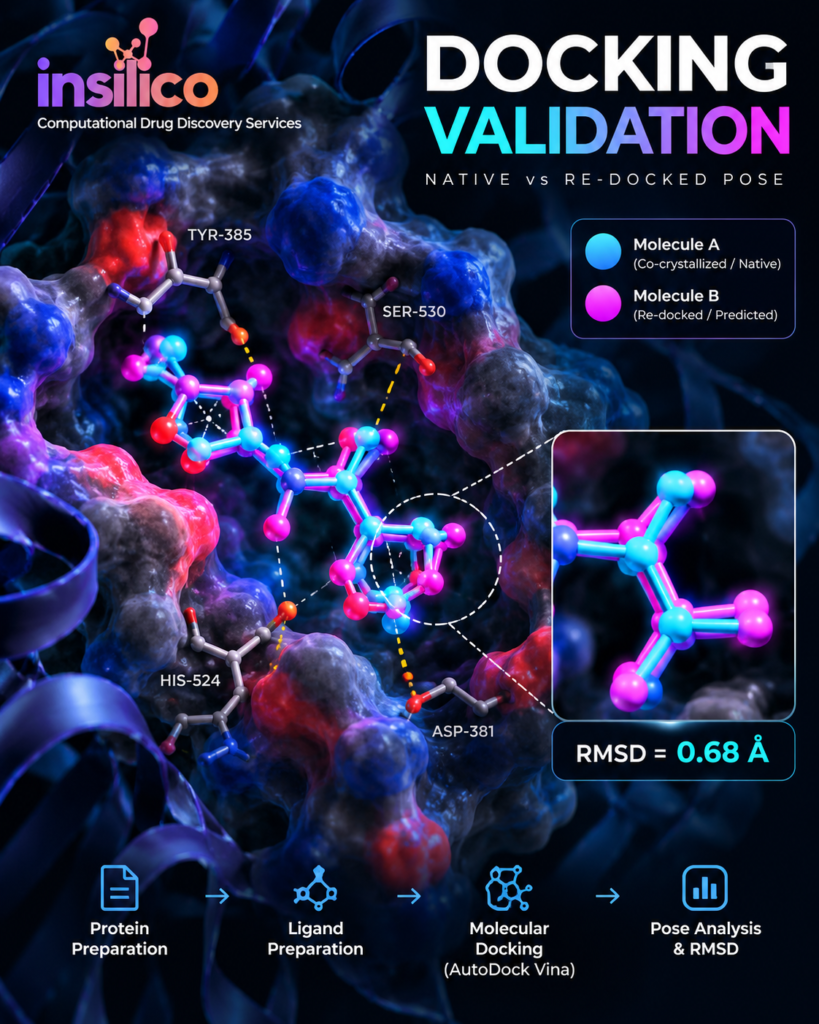
Molecular Docking Workflow: From Protein Preparation to Publication-Ready Results
Molecular docking is widely used in computational drug discovery to investigate how small molecules may interact with biological macromolecules.
Running AutoDock Vina or another docking program is relatively straightforward.
Designing a scientifically defensible docking workflow is not.
Protein selection, structure preparation, ligand preparation, binding-site definition, validation and interpretation can all influence the final results.
This article outlines a practical workflow from target selection to publication-ready molecular visualisation.
Step 1: Select the Biological Target
A docking project should begin with a biological question, not with docking software.
The selected protein should have a defensible relationship with the disease, pathway or mechanism under investigation.
Evidence may come from:
- experimental literature,
- pathway analysis,
- network pharmacology,
- genomic studies, or
- established pharmacological mechanisms.
Docking against an irrelevant target can generate technically correct calculations with little biological meaning.
Step 2: Select an Appropriate Protein Structure
Experimentally determined protein structures can be obtained from the Protein Data Bank (PDB).
Researchers should consider factors such as:
- experimental method,
- structural resolution,
- presence of missing residues,
- relevant chains,
- mutations,
- co-crystallized ligands, and
- whether the available structure represents an appropriate biological state.
A structure containing a relevant native ligand can be particularly valuable because it may help identify the binding site and enable redocking validation.
Step 3: Prepare the Protein
Raw protein structures are not always immediately suitable for docking.
Preparation may involve:
- selecting the appropriate chain,
- removing irrelevant molecules,
- evaluating crystallographic water molecules,
- adding hydrogens,
- assigning required charges,
- addressing structural issues where appropriate, and
- converting the structure into the format required by the docking software.
Protein preparation decisions should be documented because they can influence the predicted binding poses.
Step 4: Prepare the Ligands
Ligands must also be prepared carefully.
A typical workflow can involve:
- obtaining or drawing the molecular structure,
- checking chemical identity,
- generating an appropriate 3D structure,
- assigning protonation or charge states where required,
- performing geometry optimisation, and
- converting the ligand to the required docking format.
Reliable molecular identifiers such as PubChem CID, canonical SMILES or InChI can help maintain traceability.
Step 5: Define the Binding Site
One of the most important decisions is where docking should occur.
When a co-crystallized ligand is available, its experimental position can help define the active site.
Alternatively, binding-site information may come from:
- published literature,
- experimentally characterised catalytic residues,
- known inhibitor-binding regions, or
- carefully selected pocket-prediction approaches.
The search space should adequately cover the relevant binding region without being unnecessarily large.
Step 6: Validate the Docking Protocol
When an appropriate co-crystallized ligand is available, redocking provides a useful validation strategy.
The native ligand is removed and docked back into the binding site.
Its predicted pose is then compared with the experimental orientation.
RMSD analysis can quantify the difference between these conformations.
An RMSD around or below 2 Å is commonly used as a practical indication of successful pose reproduction, although interpretation depends on the molecular system and methodology.
Step 7: Perform Molecular Docking
Once the protocol has been prepared and, where possible, validated, the study compounds can be docked.
Software such as AutoDock Vina, often used through interfaces such as PyRx, can generate multiple predicted poses and corresponding scoring-function estimates.
Rather than automatically selecting compounds solely by the most negative score, researchers should examine the predicted binding orientation and interaction pattern.
Step 8: Analyse Protein–Ligand Interactions
Selected complexes can be visualised using tools such as PyMOL and Discovery Studio Visualizer.
Analysis may include:
- hydrogen bonds,
- hydrophobic interactions,
- π–π interactions,
- π–alkyl interactions,
- electrostatic interactions,
- interacting amino-acid residues, and
- comparison with a reference ligand.
Particular attention should be given to interactions involving residues known to contribute to the biological activity of the binding site.
Step 9: Compare With a Reference Compound
Where scientifically appropriate, including a known inhibitor, approved drug or native ligand can provide useful context.
The comparison should not rely exclusively on docking scores.
A study can instead examine:
- binding orientation,
- important residue interactions,
- interaction types,
- predicted affinity, and
- consistency with known structural biology.
Step 10: Prepare Publication-Ready Figures
Good visualisation is part of scientific communication.
Useful outputs can include:
3D binding pose
Shows the ligand within the protein-binding pocket.
2D interaction diagram
Summarises residues and interaction types.
Redocking overlay
Shows experimental and computationally predicted native-ligand poses.
Comparative docking figure
Allows important compounds and reference molecules to be evaluated side by side.
Figures should be exported at suitable resolution and labelled clearly for manuscript or thesis preparation.
Avoid Overinterpreting Docking Results
Perhaps the most important principle in molecular docking is understanding what the method cannot demonstrate.
Docking does not prove that a compound:
- binds experimentally,
- inhibits the protein,
- produces a therapeutic effect,
- is safe, or
- will function as a drug.
Instead, molecular docking generates computational hypotheses about potentially favourable binding modes and interactions.
Experimental studies remain necessary to validate biological activity.
Building a Stronger Computational Study
Docking becomes considerably more informative when integrated with complementary approaches.
For example:
ADMET analysis can evaluate predicted pharmacokinetic and toxicity characteristics.
Network pharmacology can help identify biologically relevant targets and pathways.
Molecular dynamics simulations can investigate the behaviour of selected complexes over time when appropriate.
Together, these methods can provide a more comprehensive computational framework than docking scores alone.
Final Thoughts
Reliable molecular docking is not simply a matter of uploading a protein and ligand and recording the lowest binding score.
A strong workflow requires appropriate target selection, careful structural preparation, defensible binding-site selection, protocol validation, interaction analysis and cautious biological interpretation.
When these steps are documented transparently, molecular docking becomes a much more useful tool for generating hypotheses and prioritising compounds for further investigation.
Need help with molecular docking analysis?
InSilicoLogics provides protein and ligand preparation, AutoDock Vina docking, redocking and RMSD validation, protein–ligand interaction analysis, comparative docking and publication-ready figures for academic research.